
What we do
LABNETWORK PATHOLOGY AND MEDICAL LABORATORY is uniquely positioned and has the expertise to provide a full range of diagnostic testing.
Please click on the items below to reveal the details

Labnetwork offers the best scanning experience and technology in Ultrasound Imaging. With an array of specialists and Consultant radiologists, we serve as a reference center to other ultrasound centers in Nigeria
Our Ultrasound Services Include:
The 4D Advantage

This is also referred to as laboratory medicine. Clinical pathology concerns the analysis of blood, urine and tissue samples to examine and diagnose disease. Examples of the information clinical pathology laboratories may provide include blood count, blood clotting and electrolyte results. A clinical pathologist is usually trained in microbiology, hematology or blood banking, but not at the same expert level as someone who specializes in one of these fields. Labnetwork has staff that specialize in the different fields of Clinical Pathology, for more accuracy and reliability
The fields under Clinical Pathology include:
Special Pathology
Labnetwork also delivers accurate timely results for special investigations such as Viral loads (For viral diseases) and DNA Paternity etc

With an in-house consultant pathologist, we deliver accurate histopathology results at the fastest possible time
This discipline can be subdivided into several disciplines and examples of these are given below:
- Histology – Samples of bodily tissues and organs are prepared and examined in order to detect and diagnose disease. The architecture of tissue is observed at a microscopic level and the relationship between different cell and tissue types is examined.
- Cytology – Bodily fluids and tissues are examined at the cellular level in order to screen for and diagnose disease and help aid treatment decisions. A cytologist will examine how cells look, form and function.
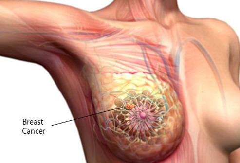
With regular screenings and follow-up care, breast and cervical cancers can be stopped before they start or found early when treatment works best. Getting screened regularly is important because women who are not screened or have not been screened in a long time could have cancer and not know it.
Getting screened for Breast Cancer
A mammogram is a low-dose X-ray of the breast. Doctors use mammograms to look for early signs of breast cancer. Regular mammograms are the best test doctors have to find breast cancer early. Having regular mammograms can lower the risk of dying from breast cancer.
What to expect from a mammogram
During a mammogram, you will stand in front of a special X-ray machine. A doctor will place your breast on a clear plastic plate. Another plate will firmly press your breast from above. The plates will flatten the breast, holding it still while the X-ray is being taken. Having a mammogram is a bit uncomfortable for most women. Some women even find it painful, especially if their breasts are sensitive. But, it only takes only a few moments, and then the discomfort is over.
When to get screened
How often you get screened for breast cancer depends on many factors, including your age, your family history, and your screening history. All women ages 50 to 74 should have mammograms every 2 years. Some women might start mammograms earlier or have mammograms more frequently. Ask your doctor when and how often you should get screened and make your appointment!
Getting screened for Cervical Cancer
A Pap test (also called a Pap smear) checks for changes in the cells of your cervix that could turn into cancer over time. The Pap test can find these cells early when treatment works best.
What to expect from a Pap test
During a Pap test, you will be asked to lie on your back with your feet in stirrups. The doctor will use a plastic or metal instrument, called a speculum, to widen the vagina. This helps the doctor examine the vagina and the cervix, and collect cells and mucus from the cervix and the area around it. The cells will then be placed on a slide or in a bottle of liquid and sent to a laboratory. The laboratory will check to be sure that the cells are normal.
When to get screened
All women ages 21 to 65 should get screened for cervical cancer. Women who may no longer be having sex or who may feel too old to have a child should still have regular Pap tests. Cervical cancer is most often found in women who have not been screened with the Pap test in more than five years or have never been screened at all. How often you get screened for cervical cancer depends on many factors, including your age, your screening history, and your sexual activity. Talk with your doctor about when and how often you should get screened. Your doctor might be able to do a Pap test during a regular appointment, or you might have to schedule one.
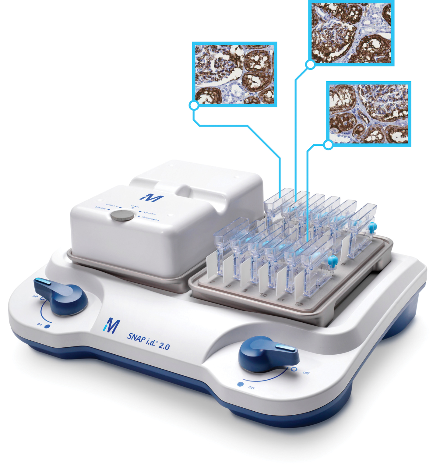
This is one of our major processes at LABNETWORK. Immunohistochemistry (IHC) refers to the process of selectively imaging antigens(e.g. proteins) in cells of a tissue section by exploiting the principle of antibodies binding specifically to antigens in biological tissues. IHC takes its name from the roots "immuno", in reference to antibodies used in the procedure, and "histo," meaning tissue (compare to immunocytochemistry). The procedure was conceptualized and first implemented by Albert Coons in 1941.
Immunohistochemical staining is widely used in the diagnosis of abnormal cells such as those found in cancerous tumors. Specific molecular markers are characteristic of particular cellular events such as proliferation or cell death (apoptosis). Immunohistochemistry is also widely used in basic research to understand the distribution and localization of biomarkers and differentially expressed proteins in different parts of a biological tissue.
Visualising an antibody-antigen interaction can be accomplished in a number of ways. In the most common instance, an antibody is conjugated to an enzyme, such asperoxidase, that can catalyse a colour-producing reaction (see immunoperoxidase staining). Alternatively, the antibody can also be tagged to a fluorophore, such as fluorescein or rhodamine

We deliver accurate autopsy report within the shortest possible time.
An autopsy is a thorough medical exam of a body after death. It may be done to learn about a disease or injury. Or it may be done to find out how or why a person has died.
An autopsy is done by a doctor called a pathologist. This type of doctor is an expert in examining body tissues and fluids.
Family members may ask for an autopsy to be done after a loved one has died. This is called a requested autopsy. Sometimes an autopsy is required by law. This is called a required autopsy.

The general medical examination is a common form of preventive medicine involving visits to LABNETWORK by well feeling adults on a regular basis. This is generally yearly or less frequently. It is known under several other names, such as the periodic health evaluation, annual physical, comprehensive medical exam, general health check, or preventive health examination. Most companies and organizations demand that their staff undergo these tests for work efficiency.